
AI for Workflow Optimization: A Technologist's Perspective
While much of the discussion around AI in radiology focuses on assisting radiologists with image interpretation, some of the most exciting and immediate applications of AI are designed to improve the daily workflow of the technologist. These tools aim to automate repetitive tasks, improve consistency, and reduce errors, ultimately freeing up the technologist to focus more on direct patient care.
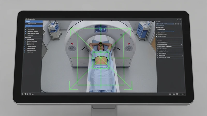
1. AI-Powered Patient Positioning
Correctly positioning a patient for a scan is a critical skill that directly impacts image quality. New AI-powered systems use cameras integrated into the scanner to streamline this process.
- How it Works: The AI uses a camera to recognize the patient's body on the scanner table. It automatically identifies anatomical landmarks and suggests the precise table position and centering needed for the exam.
- The Benefit: This can significantly reduce the time needed for setup, especially for less experienced technologists. It ensures a high degree of consistency and accuracy, reducing the need for repeat scans due to positioning errors.
2. Automated Scan Protocoling
Choosing the correct scan protocol from a long list can be complex. AI can help automate and optimize this decision-making process.
- How it Works: The AI algorithm can read the exam description from the RIS (e.g., "CT Abdomen/Pelvis with contrast for liver lesion"). Based on this text, and potentially the patient's prior imaging history, it can automatically select and load the most appropriate scan protocol.
- The Benefit: This reduces the chance of human error in selecting the wrong protocol and improves the overall standardization of care across an institution. It also speeds up the time between patients.
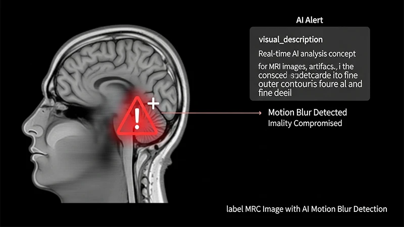
3. Real-Time Image Quality Control
One of the biggest challenges for a technologist is knowing whether an image is of diagnostic quality before the patient leaves the scanner, especially in MRI where scans are long.
- How it Works: An AI algorithm can analyze images in real-time as they are acquired. It can immediately detect common artifacts, such as patient motion.
- The Benefit: If the AI detects a motion-corrupted sequence, it can alert the technologist instantly. This allows the technologist to repeat that specific part of the scan while the patient is still on the table, rather than discovering the problem later and having to call the patient back for another appointment.
4. AI-Driven Dose Optimization
For CT scans, AI plays a crucial role in adhering to the ALARA principle. Modern reconstruction algorithms use AI techniques to produce high-quality images from scans performed at a much lower radiation dose.
- How it Works: Traditional reconstruction methods required a certain amount of radiation to create a clear, "low-noise" image. AI-based iterative reconstruction algorithms are much better at cleaning up the "noise" from low-dose scans.
- The Benefit: This allows technologists to confidently scan patients at a significantly reduced radiation dose without sacrificing the diagnostic quality needed by the radiologist.
Conclusion: The Technologist's Smart Co-Pilot
The integration of AI into the technologist's workflow is not about replacing human skill but augmenting it. These tools act as a "smart co-pilot," handling the routine, repetitive, and data-heavy tasks. This automation leads to greater efficiency, improved consistency, and enhanced patient safety. By offloading these cognitive burdens, AI empowers the technologist to dedicate more of their time and expertise to what they do best: ensuring the comfort, safety, and well-being of the patient.
Comments