
Learning Hub
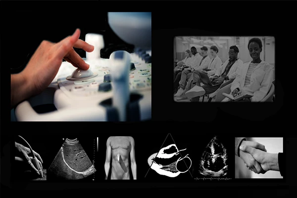
Medical Imaging Glossary
Confused by a term? Find clear definitions for common words and acronyms in radiology.
What is a DICOM File? A Complete Guide
A deep dive into the DICOM format. Understand what a .dcm file is, how it stores medical data, and its importance in modern medicine.
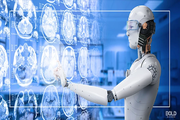
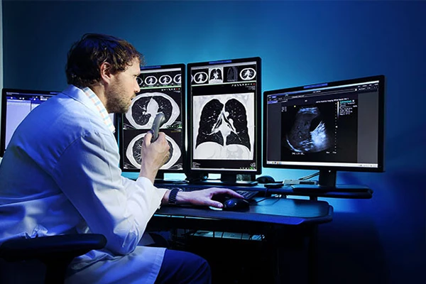
What is Artificial Intelligence (AI) in Radiology? A Foundational Guide
Explaining machine learning, deep learning, and how algorithms are trained to analyze medical images.

Will AI Replace Radiologists and Technologists? An Honest Look
An analysis of why AI is more likely to augment, not replace, radiologists and technologists.
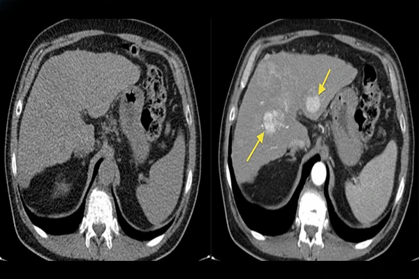
How AI is Helping to Read Chest X-Rays and Mammograms
A look at practical AI applications like triage and computer-aided detection in high-volume studies.
AI for Workflow Optimization: A Technologist's Perspective
An look at how AI is being used to improve the workflow for radiology technologists.
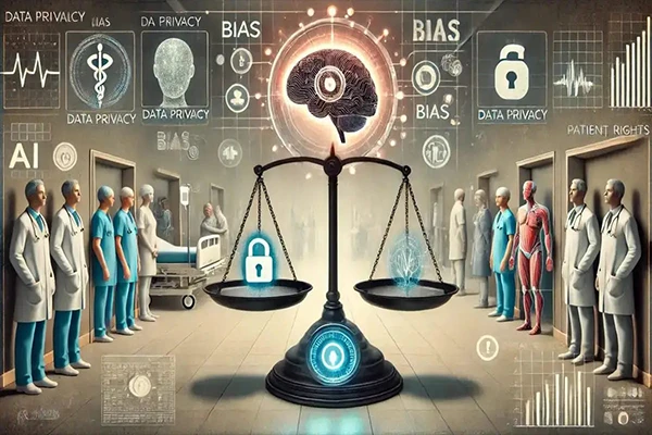
The Ethical Challenges and Biases of AI in Medical Imaging
An exploration of algorithmic bias from training data, data privacy, and accountability for errors.
The Future of AI in Radiology: What to Expect in the Next 5 Years
A look at near-future applications of AI, including predictive analytics and automated reporting.
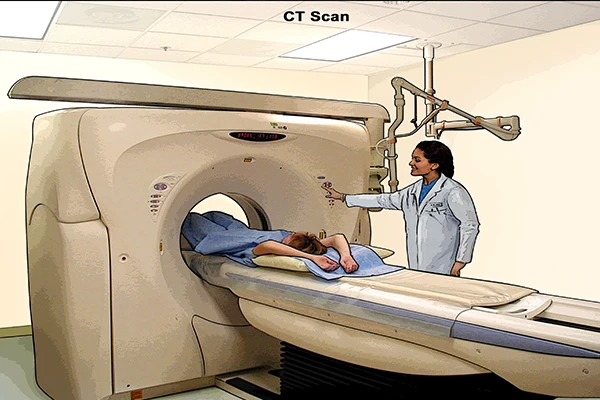
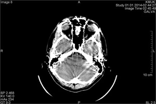
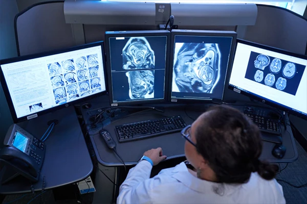
An Introduction to Computed Tomography (CT)
Learn how CT scanners use X-rays and computer processing to create detailed cross-sectional images.
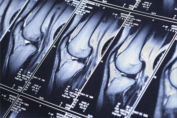
The Principles of Magnetic Resonance Imaging (MRI)
Explore how MRI uses powerful magnets and radio waves to generate sophisticated images of soft tissues.
An Introduction to Ultrasound: Seeing with Sound Waves
Learn how ultrasound imaging works using sound waves and its common applications like obstetrics.
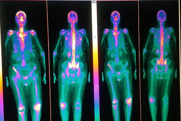
Nuclear Medicine Explained: An Overview of PET and SPECT
Understand how radiotracers are used to image physiological function with PET and SPECT scans.
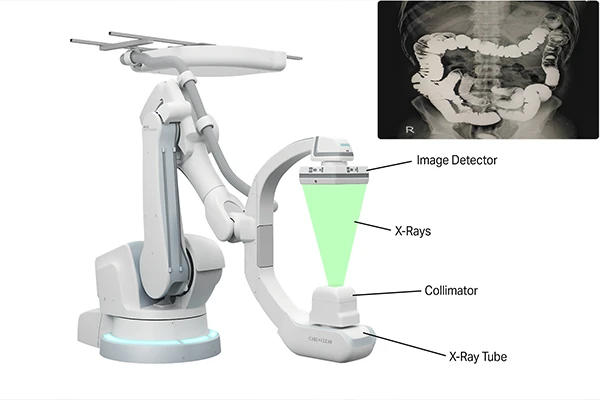
A Guide to Fluoroscopy: Visualizing the Body in Real-Time
Learn about the 'live X-ray movie' technique used for procedures like barium swallows and angiography.
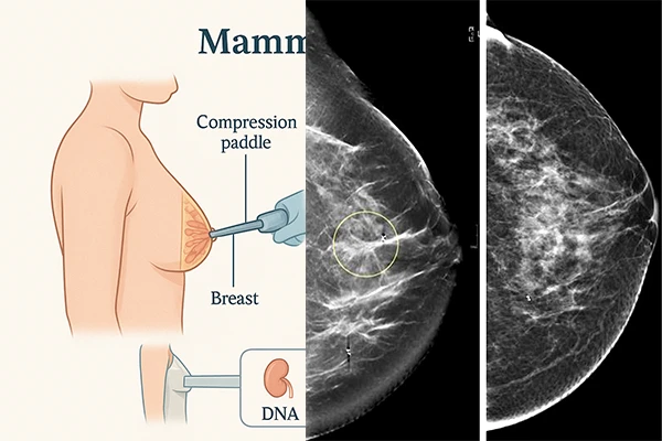
Mammography: The Gold Standard in Breast Imaging
Learn about mammography, the role of compression, and advances like 3D tomosynthesis.
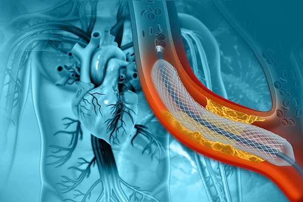
An Introduction to Interventional Radiology: Image-Guided Procedures
Learn how physicians use medical imaging to perform minimally invasive procedures and treatments.
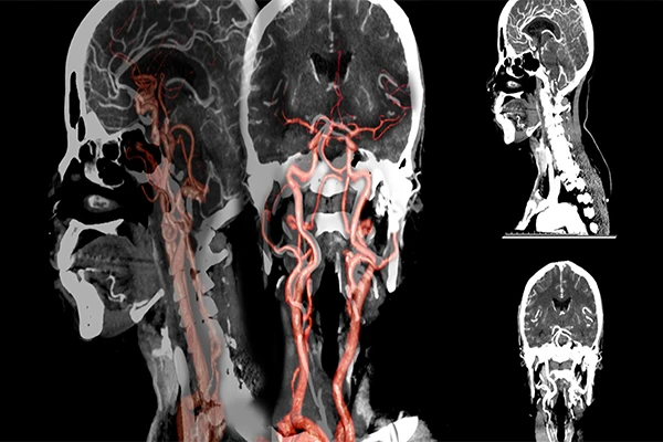
A Guide to CT Angiography (CTA): Visualizing Blood Vessels
Learn how CTA uses IV contrast and precise timing to create detailed 3D images of arteries and veins.
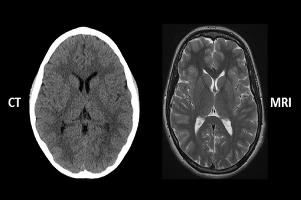
CT vs. MRI: A Detailed Comparison for Technologists
An in-depth comparison of the physics, speed, cost, and clinical applications for each modality.
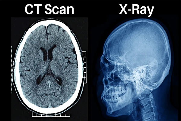
X-ray vs. CT Scan: What's the Difference?
A simple guide for patients explaining the key differences between a standard X-ray and a CT scan.
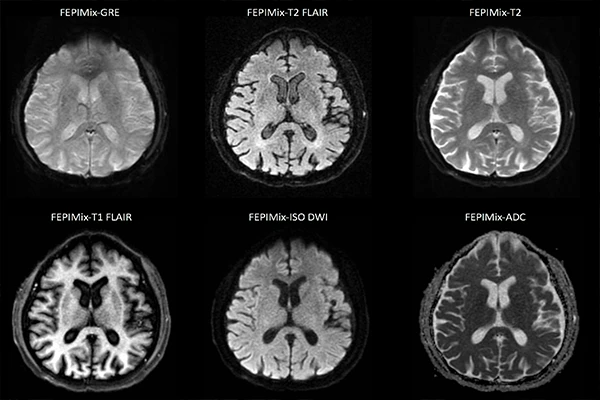
Understanding MRI Sequences: A Guide to T1, T2, FLAIR, and DWI
A deep dive into common MRI sequences, what tissues they highlight, and their clinical uses.

The Role of Contrast Agents in Radiology: A Practical Guide
Learn about iodine, gadolinium, and barium-based contrast agents, including their uses and safety.
Common Imaging Artifacts (CT & MRI) and How to Troubleshoot Them
A technologist's guide to recognizing and preventing common artifacts like motion, metal, and aliasing.
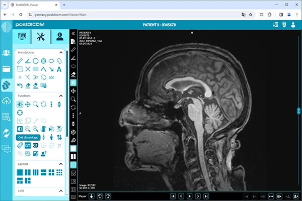
An Introduction to PACS and RIS: The IT Backbone of Radiology
Learn the difference between PACS for image storage and RIS for workflow management.
DICOM Tags Explained: A Deep Dive into Medical Image Metadata
Learn about the hidden metadata in every medical image that stores patient and study information.

DICOM vs. NIfTI: Understanding the Key Differences for Neuroimaging
A comparison of why DICOM is the clinical standard and NIfTI is preferred for academic research.

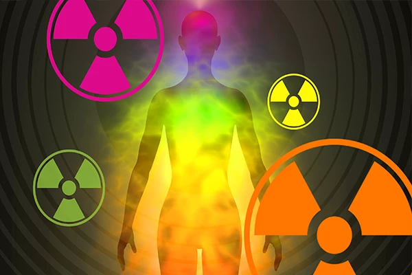
The ALARA Principle: A Practical Guide to Radiation Safety
A guide to the ALARA principle, covering the three cardinal rules: Time, Distance, and Shielding.
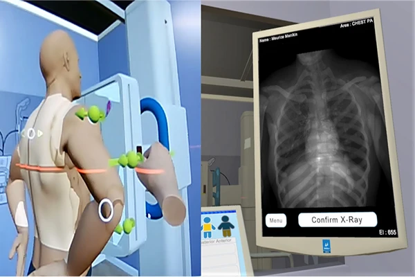
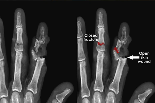
Patient Positioning in Radiography: A Visual Guide
A guide to positioning for common projections like chest, hand, and foot X-rays.
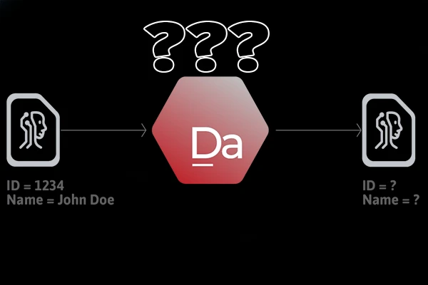
How to Anonymize DICOM Files for Research and Teaching
A practical guide on removing identifying tags and burning-in annotations to protect privacy.
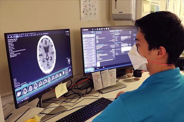
A Day in the Life of a Radiology Technologist
An inside look at the daily responsibilities, from quality control to patient care to image acquisition.
Career Paths & Specializations in Radiologic Technology
Explore career paths beyond general X-ray, including CT, MRI, Interventional Radiology, and more.
Why Do I Need an IV Contrast for My Scan? (CT & MRI)
A patient's guide on why IV contrast is used to highlight blood vessels, organs, and abnormalities.
How to Read Your Radiology Report: A Guide to Common Terms
A patient-friendly guide to understanding your radiology report and its common terms.

Preparing for Your Medical Imaging Exam: A Checklist for Patients
A simple checklist for patients on how to prepare for exams like CT, MRI, and Ultrasound.
What is a "STAT" Read in Radiology? The Race Against Time
An explanation of what a 'STAT' read means and how the workflow is prioritized for critical findings.
Understanding Radiation Dose: mSv, Gray, and Dose Reports
A guide to the units of radiation measurement, including absorbed dose (Gray) and effective dose (Sievert).
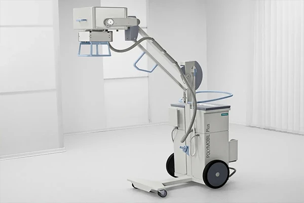
A Technologist's Guide to Portable X-ray
A guide to portable radiography, covering its use cases and unique technical and safety challenges.
The Role of Radiology in Trauma: The Primary Survey
An overview of how imaging is used in the critical 'primary survey' of a trauma patient.
Research Topics Coming Soon
This section will feature in-depth research topics and case studies in the field of medical imaging. Please check back later for updates!