
The Role of Radiology in Trauma: The Primary Survey
In the high-stakes environment of a trauma bay, time is the most critical factor. The immediate goal is to identify and treat life-threatening injuries, a process guided by the Advanced Trauma Life Support (ATLS) protocol. Medical imaging, particularly portable X-ray and rapid CT scanning, plays an indispensable role in this "primary survey," helping the trauma team see inside the body to make split-second, life-saving decisions.
The Initial Assessment: Portable X-ray
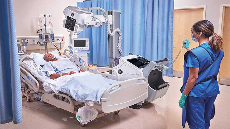
Often, before the patient is stable enough to move to a CT scanner, the first images are acquired directly in the trauma bay using a portable X-ray machine. The two most common and critical portable exams are:
- AP Chest X-ray: This is crucial for quickly identifying immediate threats to breathing and circulation, such as a collapsed lung (pneumothorax), significant fluid or blood in the chest (hemothorax), or a widened mediastinum that could suggest a major aortic injury.
- AP Pelvis X-ray: A fractured pelvis can be a source of massive internal bleeding. A simple AP pelvis X-ray can immediately identify a severe fracture, alerting the team to the risk of hemodynamic instability.
These initial images are not meant to be perfect; they are rapid screening tools to rule in or rule out the most immediate life threats.
The Gold Standard: The "Pan-Scan" CT
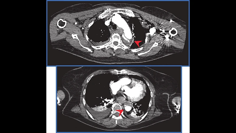
Once the patient is stabilized enough for transport, the definitive imaging study is typically a "pan-scan" or "trauma scan." This involves a rapid CT scan from the head to the pelvis, usually after an injection of IV contrast. The speed of a modern multi-detector CT scanner is a game-changer, allowing this entire process to be completed in just a few minutes.
The pan-scan provides a comprehensive evaluation of potential injuries:
- Head CT: Identifies intracranial bleeding, skull fractures, and brain swelling.
- C-Spine CT: Clears the cervical spine of any fractures or ligamentous injuries, which is critical before removing a neck collar.
- Chest CT: Provides a much more detailed view than an X-ray, able to detect subtle lung contusions, small pneumothoraces, and injuries to the aorta and other major vessels.
- Abdomen/Pelvis CT: The most important part for identifying internal injuries. It can show lacerations to solid organs like the liver, spleen, and kidneys, as well as signs of active bleeding ("contrast extravasation").
FAST Scan (Focused Assessment with Sonography for Trauma)
In some trauma centers, a portable ultrasound machine is used at the bedside as part of the initial assessment. The FAST scan is a quick, non-invasive exam where the physician looks for the presence of free fluid (which usually indicates blood) in key areas of the abdomen and around the heart. A positive FAST scan can be an indication to take the patient directly to the operating room.
Conclusion: Guiding Critical Care
Radiology is the eyes of the trauma team. From the initial portable X-rays in the bay to the comprehensive detail of the pan-scan CT, medical imaging provides the crucial information needed to diagnose and prioritize injuries. This rapid, precise visualization of internal trauma is a cornerstone of modern emergency medicine and is directly responsible for improving patient outcomes and saving lives in the critical moments after a severe injury.
Comments