
An Introduction to Interventional Radiology: Image-Guided Procedures
Interventional Radiology (IR) is a dynamic and rapidly growing subspecialty of radiology that uses medical imaging to perform minimally invasive procedures. Often described as "surgery through a pinhole," IR represents a significant shift from traditional open surgery. Interventional radiologists are physicians who use tools like catheters, wires, and needles, guided by imaging technologies like fluoroscopy, CT, and ultrasound, to diagnose and treat a wide range of conditions.
The Core Concept: Seeing is Guiding
The fundamental principle of IR is to use the detailed "road map" provided by medical imaging to navigate instruments through the body to a target area without making large incisions. A tiny nick in the skin is often all that is needed to access blood vessels or organs. This approach offers numerous patient benefits, including reduced risk, less pain, and significantly faster recovery times compared to conventional surgery.
The Tools of the Trade
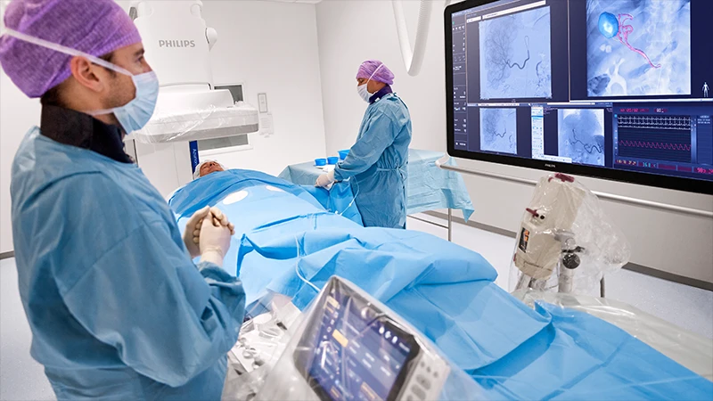
An interventional suite is a hybrid of an operating room and an advanced imaging room. The team, which includes the interventional radiologist, a specialized technologist, and a nurse, uses a variety of tools:
- Imaging Guidance: Fluoroscopy (live X-ray) is the most common tool, allowing for real-time visualization of instruments. CT and Ultrasound are also used for guiding needles and catheters with precision.
- Catheters and Wires: Thin, flexible tubes (catheters) and wires are skillfully navigated through arteries and veins to reach virtually any part of the body.
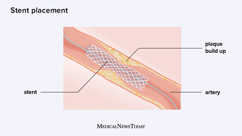
- Specialized Devices: Through these catheters, radiologists can deliver tiny tools like stents (to open blocked vessels), coils (to block bleeding), and ablation probes (to destroy tumors).
Common Interventional Procedures
The scope of IR is vast and constantly expanding. Some of the most common procedures include:
- Angiography and Angioplasty/Stenting: Visualizing blood vessels with contrast and then using balloons (angioplasty) or metal scaffolds (stents) to open blockages. This is commonly done in the legs, kidneys, and carotid arteries.
- Embolization: A procedure to block blood flow to a specific area. This can be used to stop bleeding (e.g., after trauma), cut off the blood supply to a tumor, or treat conditions like uterine fibroids.
- Thrombectomy: The emergency removal of a blood clot, most critically from the brain during an acute stroke.
- Biopsies and Drainages: Using CT or ultrasound guidance to precisely place a needle to obtain a tissue sample (biopsy) or to drain an abnormal fluid collection (like an abscess).
- Tumor Ablation: Using probes that deliver heat (radiofrequency ablation) or cold (cryoablation) to destroy cancer cells in organs like the liver, kidney, or lung.
The Role of the IR Technologist
The technologist in an IR suite is a highly skilled professional who acts as the radiologist's essential partner. They are responsible for operating the complex imaging equipment, managing radiation safety for everyone in the room, and anticipating the needs of the procedure by preparing and handing off the correct wires, catheters, and devices.
Conclusion: The Future of Modern Medicine
Interventional Radiology has revolutionized medicine by offering less invasive, lower-risk, and highly effective alternatives to traditional surgery. By combining the diagnostic power of imaging with targeted therapeutic procedures, IR specialists provide cutting-edge care that helps patients recover faster and live longer. It is one of the most innovative and rapidly evolving fields in all of medicine.
Comments