
How AI is Helping to Read Chest X-Rays and Mammograms
Chest X-rays and screening mammograms are two of the highest-volume studies in any radiology department. Radiologists must interpret hundreds of these images every day, looking for subtle abnormalities in a sea of normal cases. This high-volume, repetitive work is a perfect area for Artificial Intelligence to provide valuable assistance, acting as a tireless and vigilant partner to the human expert.
The Challenge: Finding the Needle in the Haystack
The primary challenge in reading screening studies is perceptual error. A small lung nodule or a tiny cluster of microcalcifications can be easily missed, especially when a radiologist is reading their 200th case of the day. This is where AI excels. An AI algorithm doesn't get tired or distracted. It can analyze every pixel of every image with the same level of scrutiny.
Application 1: Computer-Aided Detection (CADe)
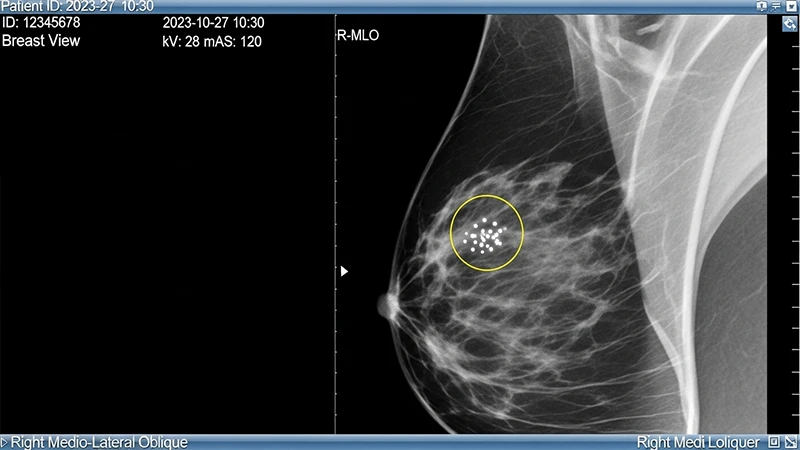
This is one of the earliest and most common applications of AI in imaging. A Computer-Aided Detection (or CADe) algorithm analyzes an image and places markers or circles on areas that it identifies as potentially abnormal. It acts as a "spell-checker" for the radiologist.
- In Mammography: CADe systems are trained to detect suspicious masses and clusters of microcalcifications, which can be an early sign of breast cancer.
- In Chest X-rays: AI can highlight potential lung nodules, areas of pneumonia, or a collapsed lung (pneumothorax).
The radiologist then reviews these marks. They use their expertise to decide if the AI's finding is a true abnormality or a "false positive" (e.g., normal overlapping blood vessels). The final diagnosis is always made by the human.
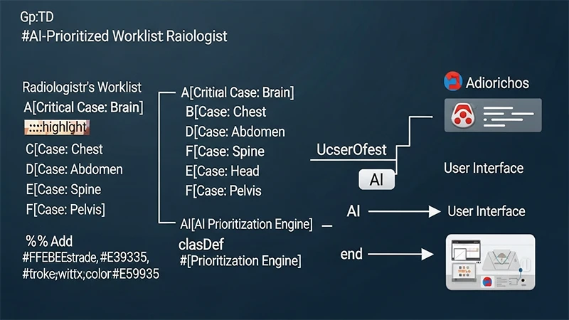
Application 2: Workflow Triage
In a busy hospital, a radiologist's worklist can be hundreds of studies long. Some of those cases may contain critical, time-sensitive findings, but they might be buried at the bottom of the list. Triage AI helps solve this problem.
The AI algorithm analyzes every study as soon as it arrives in the PACS. If it detects a finding that requires immediate attention—like a brain bleed on a head CT or a large pulmonary embolism—it automatically flags the study and moves it to the top of the reading queue. This ensures that the most critical cases are always seen first, which can dramatically improve patient outcomes.
Application 3: Quantitation and Measurement
AI can also automate tedious measurement tasks, improving both speed and consistency.
- In Mammography: AI can automatically measure breast density, which is an important risk factor for cancer.
- In Chest X-rays: Algorithms can measure the cardiothoracic ratio (a way to estimate heart size) or automatically track the size of a lung nodule over time on multiple follow-up scans.
By automating these tasks, AI allows radiologists to spend more of their time on complex diagnostic reasoning.
Conclusion: A Powerful Collaboration
The integration of AI into high-volume reading workflows is a prime example of human-computer collaboration. The AI provides an tireless, quantitative analysis that highlights potential areas of concern, while the radiologist provides the essential clinical context, expert judgment, and diagnostic wisdom to make the final interpretation. This partnership promises a future where medical imaging is not only faster and more efficient, but also more accurate and reliable, ultimately benefiting the patient.
Comments