
The Role of Contrast Agents in Radiology: A Practical Guide
In medical imaging, contrast agents—often called "contrast media" or "dyes"—are special substances used to improve the visibility of internal body structures. Many soft tissues have similar densities and can be difficult to differentiate on a standard scan. Contrast agents temporarily change the way imaging systems interact with the body, highlighting specific organs, blood vessels, or tissues and providing a wealth of diagnostic information.
Iodine-Based Contrast for CT and Fluoroscopy
Iodine is a chemical element that is radiopaque, meaning it effectively blocks X-rays. Because of this property, iodine-based contrast agents are widely used in CT and fluoroscopy.
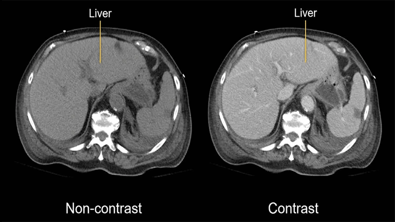
- How it Works: When injected into a vein, the iodine-rich fluid travels through the bloodstream, causing blood vessels and highly vascular organs (like the liver and kidneys) to appear bright white on a CT scan. This allows for detailed evaluation of arteries and veins (CT Angiography) and helps identify abnormalities like tumors, which often have a different blood supply than normal tissue.
- Administration: It can be given intravenously (IV), orally to highlight the GI tract, or directly into other body cavities.
Gadolinium-Based Contrast for MRI
Gadolinium is a rare earth metal with paramagnetic properties. This means it alters the magnetic field in its immediate vicinity, which can be detected by an MRI scanner.
- How it Works: Gadolinium-based contrast agents (GBCAs) are injected intravenously. They work by shortening the T1 relaxation time of nearby protons. This causes tissues where the contrast has accumulated to appear much brighter on T1-weighted images.
- Applications: It is invaluable for detecting inflammation, infection, and tumors, as these processes often cause a breakdown of the normal blood-brain barrier or have increased blood flow, allowing the gadolinium to accumulate.
Barium Sulfate for Fluoroscopy
Barium sulfate is a dense, metallic compound that is also radiopaque. It is used exclusively for imaging the gastrointestinal (GI) tract.
- How it Works: Barium is not absorbed by the body. It is administered as a liquid suspension that the patient drinks (for an upper GI study) or as an enema (for a lower GI study). It coats the lining of the esophagus, stomach, and intestines, allowing their shape and function to be visualized in real-time with fluoroscopy.
Safety of Contrast Agents
While generally very safe, all contrast agents carry some risks. Patient screening is a critical part of a technologist's job before any contrast-enhanced study.
- Iodine & Gadolinium: The primary concerns are allergic-like reactions and the effect on kidney function. Patients are always screened for a history of kidney disease, as the kidneys are responsible for clearing these agents from the body. A blood test to check creatinine levels is often required.
- Barium: The main risk is aspiration (inhaling the liquid into the lungs) in patients with swallowing difficulties, or leakage into the abdominal cavity if there is a suspected perforation in the bowel.
Patients are often asked about allergies and their medical history, and it's vital they provide accurate information. For IV contrast, patients may experience a temporary warm flushing sensation, which is a normal side effect.
Conclusion: Enhancing the Diagnostic Picture
Contrast agents are not just an add-on; they are an essential part of many diagnostic imaging procedures. By making the invisible visible, they provide crucial information that helps radiologists detect disease, characterize abnormalities, and guide treatment with greater confidence. Understanding the different types of contrast and their specific uses is fundamental to appreciating the full capability of modern radiology.
Comments