
An Introduction to Ultrasound: Seeing with Sound Waves
Ultrasound, also known as sonography, is a widely used medical imaging technique that uses high-frequency sound waves to create real-time images of the body's internal organs and structures. Unlike X-ray or CT, it does not use any ionizing radiation, making it an extremely safe and versatile tool, especially in sensitive applications like obstetrics.
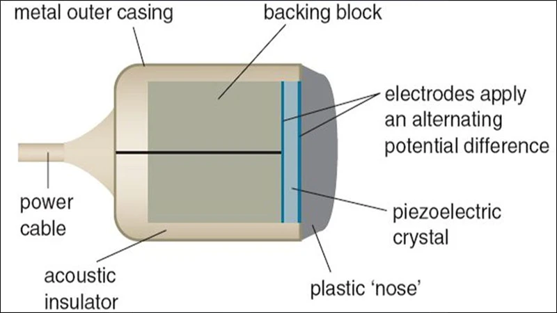
How Ultrasound Works: The Piezoelectric Effect
The core of an ultrasound machine is a small handheld device called a transducer, or probe. Inside the transducer are special crystals that have a unique property known as the piezoelectric effect.
- Generating Sound: When an electric current is applied to these crystals, they vibrate rapidly and produce sound waves at a frequency far too high for humans to hear (ultrasound).
- Listening for Echoes: These sound waves travel from the probe into the body. When they hit a boundary between different types of tissue (like between fluid and an organ), some waves are reflected back to the probe as an echo.
- Creating an Image: When the returning echo strikes the piezoelectric crystals, they convert the sound energy back into an electric signal. A computer measures the time it took for the echoes to return and their strength, then uses this information to construct a two-dimensional image on the screen.
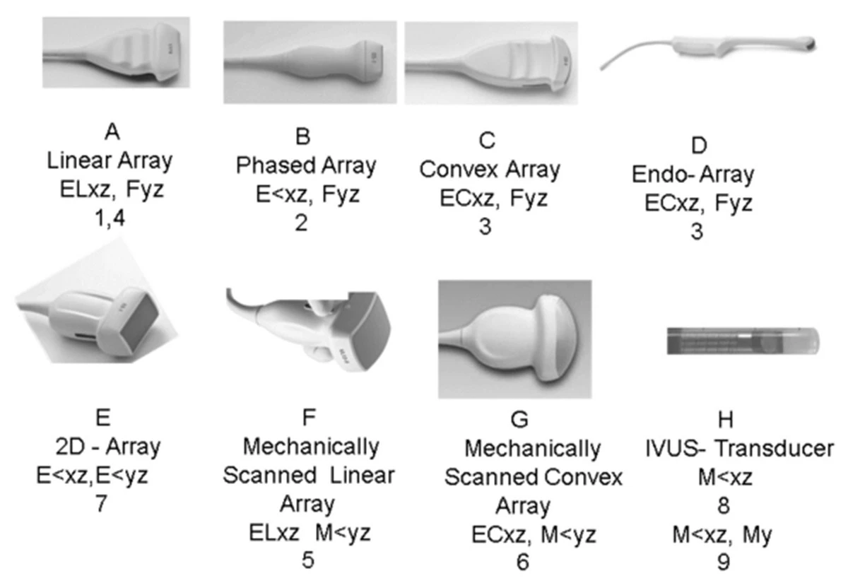
The Importance of the Transducer
Different transducers are designed for different parts of the body. The shape of the probe determines the shape of the image, and the frequency of the sound waves determines the depth and resolution.
- Linear Transducer: High frequency, provides excellent resolution for shallow structures like blood vessels and muscles.
- Curvilinear (Convex) Transducer: Low frequency, allows for deeper penetration to visualize abdominal organs like the liver and kidneys.
- Phased Array Transducer: Small footprint, ideal for imaging between the ribs to see the heart (echocardiography).
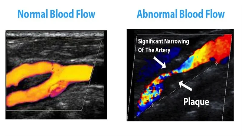
Beyond the Image: Doppler Ultrasound
One of the most powerful features of ultrasound is its ability to visualize blood flow using the Doppler effect. This is the same principle that makes a siren sound higher-pitched as it approaches you and lower-pitched as it moves away. By analyzing the frequency shift of echoes returning from moving red blood cells, the ultrasound machine can determine the speed and direction of blood flow. This is critical for diagnosing conditions like blood clots (DVT) and narrowed arteries.
Common Applications of Ultrasound
Ultrasound's safety and real-time capabilities make it invaluable in many fields:
- Obstetrics and Gynecology: Monitoring fetal growth and development, examining the uterus and ovaries.
- Cardiology: Assessing the heart's structure and function (echocardiogram).
- Abdominal Imaging: Evaluating the gallbladder, liver, kidneys, pancreas, and spleen.
- Vascular Imaging: Detecting blockages or abnormalities in arteries and veins.
- Point-of-Care (POC): Used in emergency rooms and ICUs for quick diagnostic assessments.
Conclusion: Safe, Real-Time Imaging
Ultrasound is a cornerstone of modern medical imaging due to its unique combination of safety, affordability, and real-time visualization. By harnessing the simple physics of sound waves and echoes, it provides clinicians with a dynamic and powerful tool for diagnosing a wide array of conditions without any risk of radiation exposure to the patient.
Comments