
A Guide to Fluoroscopy: Visualizing the Body in Real-Time
If a standard X-ray is like a photograph, then fluoroscopy is like a video. It is a medical imaging technique that uses X-rays to obtain real-time, moving images of the body's internal structures. This "live X-ray movie" allows physicians and surgeons to observe physiological processes, guide instruments, and perform minimally invasive procedures with precision.
How Fluoroscopy Works
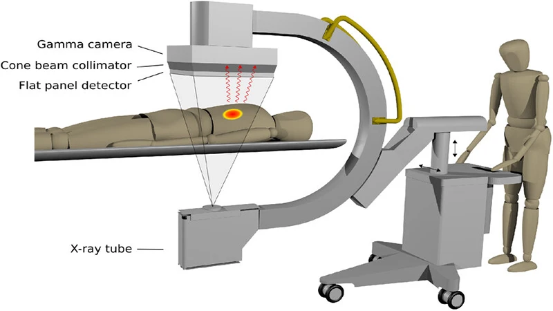
A fluoroscopy unit, often called a C-arm due to its shape, consists of an X-ray source and a detector. The patient is positioned between these two components. Unlike a standard X-ray which uses a single, short burst of radiation, fluoroscopy uses a continuous, low-dose X-ray beam that passes through the patient. The X-rays that exit the patient are captured by a detector, typically an image intensifier or a modern flat-panel detector.
The detector converts the X-ray pattern into a visible light image, which is then digitized and displayed on a monitor in the room. This allows the medical team to see the anatomy and its movement live as it happens.
The Role of Contrast Media
Many fluoroscopic procedures rely on contrast agents to make specific organs or vessels visible. Since soft tissues don't show up well on X-ray, these agents are used to coat or fill the structures of interest, making them appear white on the monitor.
- Barium: A dense, chalky liquid that is swallowed or administered as an enema to visualize the gastrointestinal (GI) tract.
- Iodine: A water-soluble contrast injected into blood vessels or joints to make them visible (angiography and arthrography).
Common Applications of Fluoroscopy
Fluoroscopy is a workhorse in many areas of medicine, particularly for guiding procedures:
- Gastrointestinal Studies: In a barium swallow or upper GI series, a radiologist watches the contrast move through the esophagus, stomach, and small intestine to diagnose swallowing problems, ulcers, or blockages.
- Interventional Radiology: Fluoroscopy is essential for guiding catheters, stents, and other devices through blood vessels in procedures like angioplasty and stenting.
- Orthopedic Surgery: Surgeons use fluoroscopy in the operating room to visualize bone alignment and ensure the correct placement of screws, plates, and joint replacements.
- Pain Management: It is used to guide the precise injection of anesthetics and steroids into specific areas of the spine or joints.
Radiation Safety: A Critical Consideration
Because fluoroscopy involves a continuous X-ray beam, radiation safety is of the utmost importance for both the patient and the medical staff. Fluoroscopy systems are designed with features to minimize the dose, such as pulsed fluoroscopy (which rapidly turns the beam on and off) and last-image hold.
The core principles of radiation safety—Time, Distance, and Shielding—are strictly followed. Technologists and physicians limit the beam-on time, increase their distance from the X-ray source whenever possible, and wear lead aprons, thyroid shields, and protective glasses.
Conclusion: Guiding Modern Medicine
Fluoroscopy is a vital imaging modality that provides a dynamic view of the body at work. Its ability to produce real-time images makes it an indispensable tool for a vast range of diagnostic studies and minimally invasive interventional procedures. While managing radiation dose is a key part of its use, fluoroscopy's guidance capabilities have revolutionized many aspects of surgery and interventional medicine, allowing for safer and more effective treatments.
Comments