
Understanding MRI Sequences: A Guide to T1, T2, FLAIR, and DWI
The power of MRI lies in its versatility. By adjusting the timing and parameters of the radiofrequency (RF) pulses and signal detection, technologists can generate a variety of "image weightings" or sequences. Each sequence is designed to highlight specific tissue characteristics, providing different pieces of the diagnostic puzzle. A standard MRI exam is not a single scan, but a collection of these specialized sequences.
Let's break down some of the most common and fundamental sequences used in neuroimaging and beyond.
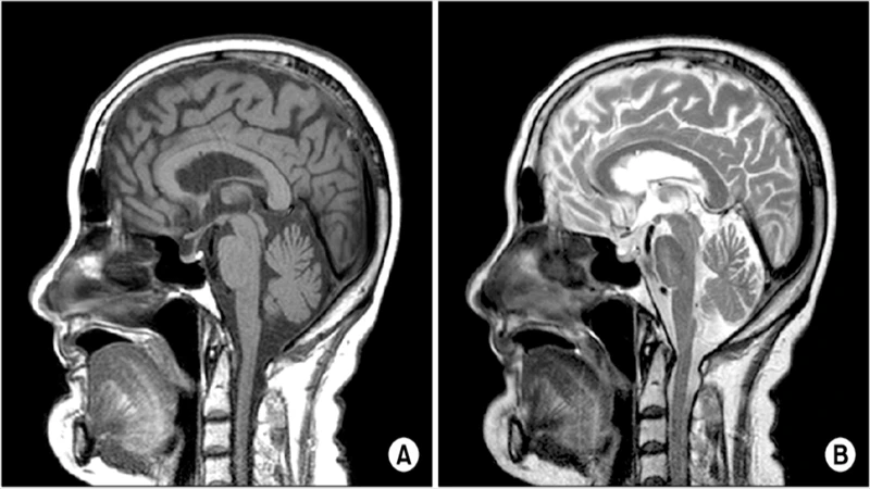
T1-Weighted (The "Anatomy" Sequence)
The T1-weighted sequence is the workhorse for visualizing normal anatomy. It provides excellent contrast between different soft tissues, making it easy to distinguish gray matter from white matter in the brain, for example.
- Key Characteristic: Fat is bright, and water (like cerebrospinal fluid, or CSF) is dark.
- Mnemonic: Think T"1" = "1" tissue type (fat) is bright.
- Common Uses: Visualizing the structure of organs, identifying anatomical landmarks. It is also the primary sequence used after the administration of gadolinium contrast, as the contrast agent appears very bright on T1 images.
T2-Weighted (The "Pathology" Sequence)
The T2-weighted sequence is extremely sensitive to changes in water content. Since most pathologies—such as tumors, inflammation, infection, and trauma—involve an increase in water (edema), they will appear bright on a T2 image.
- Key Characteristic: Water and fat are both bright.
- Mnemonic: Think T"2" = "2" tissue types (water and fat) are bright.
- Common Uses: Detecting edema, inflammation, tumors, and other disease processes. It's often the first sequence a radiologist looks at to spot an abnormality.
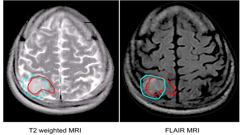
FLAIR (Fluid Attenuated Inversion Recovery)
FLAIR is a clever modification of the T2 sequence. It takes a standard T2 image and digitally "erases" the bright signal from normal fluid, like the CSF in the brain's ventricles. This makes pathology near fluid-filled spaces much easier to see.
- Key Characteristic: It's a T2 image where water/CSF is dark, but pathology (edema) remains bright.
- Common Uses: Essential in neuroimaging for detecting subtle abnormalities near the ventricles, such as the plaques seen in multiple sclerosis or subtle strokes.
DWI (Diffusion Weighted Imaging)
DWI is a highly specialized sequence that measures the random motion (diffusion) of water molecules within tissue. In healthy tissue, water molecules can move freely. However, in certain disease states, this movement is restricted.
- Key Characteristic: Tissues with restricted water diffusion appear bright.
- Primary Use: Its most critical application is in the early detection of acute ischemic stroke. Within minutes of a stroke occurring, cytotoxic edema causes brain cells to swell, restricting water diffusion. This area will appear brightly on a DWI sequence long before it's visible on T1 or T2 images. It's also used in tumor imaging.
Conclusion: A Diagnostic Toolkit
T1, T2, FLAIR, and DWI are just a few of the many sequences available in the MRI toolkit. A radiologist interprets an exam by comparing how an area of tissue appears across these different sequences. An abnormality might be dark on T1, bright on T2 and FLAIR, and show restricted diffusion on DWI. This multi-faceted view allows for a highly specific and confident diagnosis. Understanding the basic principles of these core sequences is the first step to appreciating the true diagnostic power of MRI.
Comments