
How to Read Your Radiology Report: A Guide to Common Terms
After a medical imaging exam, a radiologist will interpret your images and write a detailed report. Receiving this report can be intimidating, as it is written for your doctor and filled with technical medical terms. While it is **essential to discuss the results with your doctor**, understanding the structure and common language of the report can help you feel more informed and prepared for that conversation.
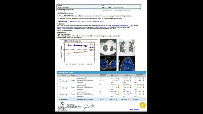
The Structure of a Radiology Report
Most reports are organized into a standard format:
- Type of Exam: States what kind of scan was performed (e.g., "CT of the Abdomen and Pelvis with IV Contrast").
- Clinical History/Indication: A brief sentence explaining why you had the scan (e.g., "Right lower quadrant abdominal pain.").
- Comparison: Lists any prior, relevant exams that the radiologist used for comparison (e.g., "Compared to CT scan from January 5, 2024.").
- Technique: Briefly describes how the scan was performed.
- Findings: This is the main body of the report. The radiologist describes, system by system, what they see on your images, commenting on each organ and structure.
- Impression: This is the most important section. It is the radiologist's summary and conclusion. If there is a key finding or diagnosis, it will be stated here.
A Glossary of Common Terms
Here are some common words and phrases you might encounter in the "Findings" and "Impression" sections:
Directional and Positional Terms
- Anterior / Posterior: Front / Back
- Superior / Inferior: Upper / Lower
- Medial / Lateral: Toward the midline / Toward the side
- Proximal / Distal: Closer to the center of the body / Farther from the center of the body (used for limbs)
Descriptive Terms for Findings
- Unremarkable / Within normal limits: This is good news! It means the radiologist saw nothing abnormal in that area.
- Lesion / Mass / Nodule: These are general terms for an abnormal area of tissue. They do not automatically mean cancer. A lesion could be a simple cyst, a scar, or a tumor (which could be benign or malignant).
- Benign / Malignant: Not cancerous / Cancerous.
- Hypodense / Hyperdense (CT): Appears darker than surrounding tissue / Appears brighter than surrounding tissue.
- Hypointense / Hyperintense (MRI): Appears darker than surrounding tissue / Appears brighter than surrounding tissue.
- Enhancement: An area that "lights up" or appears brighter after receiving an IV contrast injection. This indicates increased blood flow, which can be seen in tumors and inflammation.
- Edema: Swelling caused by an excess of fluid in the tissue.
Important Phrases in the "Impression"
- "No acute findings": A very common phrase in emergency room reports. It means there are no urgent or life-threatening problems that require immediate attention (like a bleed, major fracture, or appendicitis).
- "Correlate clinically": This is a note from the radiologist to your doctor. It means the imaging finding is non-specific, and its importance depends on your symptoms and physical exam. For example, a small amount of fluid could be normal or a sign of a problem, depending on the clinical context.
- "Recommend follow-up": The radiologist may suggest another imaging test to get a better look at a finding or to monitor it over time.
Conclusion: A Tool for Your Doctor
Your radiology report is a complex medical document, and it is just one piece of your overall health picture. It's easy to get worried or confused by the technical language. Remember that the report is a tool written for your doctor, who will interpret the findings in the context of your personal medical history, physical exam, and lab results. Always use the report as a starting point for a detailed conversation with your healthcare provider to get the full story.
Comments