
Understanding Radiation Dose: mSv, Gray, and Dose Reports
Quantifying radiation is a complex topic, and several different units are used in medical imaging to measure it. While patients often hear about dose in terms of "years of background radiation," technologists and physicists use more precise scientific units. Understanding the difference between terms like Gray and Sievert is key to appreciating the science of radiation safety.
Absorbed Dose: Gray (Gy)
The most fundamental unit is the **Absorbed Dose**, measured in **Gray (Gy)**. One Gray is defined as the absorption of one joule of radiation energy per kilogram of matter.
Think of it like this: The Gray measures the raw amount of energy that is deposited into a specific tissue. It tells you the concentration of energy absorbed by that area. In CT, you will see values like the CTDIvol (CT Dose Index) measured in milligray (mGy).
Equivalent Dose: Sievert (Sv)
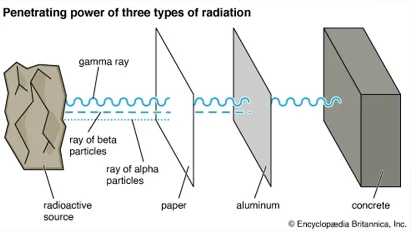
Not all types of radiation have the same biological effect. An alpha particle, for example, is much more damaging than an X-ray photon, even at the same absorbed dose. The **Equivalent Dose**, measured in **Sieverts (Sv)**, accounts for this difference.
It is calculated by taking the absorbed dose (in Gy) and multiplying it by a "radiation weighting factor." For the X-rays used in medical imaging, this factor is 1. Therefore, for our purposes, 1 Gy of absorbed dose equals 1 Sv of equivalent dose.
Effective Dose: Sievert (Sv)
Different body tissues also have different sensitivities to radiation. The reproductive organs and bone marrow, for example, are more sensitive than skin. The **Effective Dose**, also measured in **Sieverts (Sv)**, accounts for this by considering which organs were irradiated.
It is calculated by taking the equivalent dose to each organ and multiplying it by a "tissue weighting factor." The sum of these values gives the effective dose. The effective dose is the most useful value for estimating the overall, long-term health risk to a person from a given exposure. When you hear that a chest CT is "7 mSv," this is referring to the effective dose.
The CT Dose Report
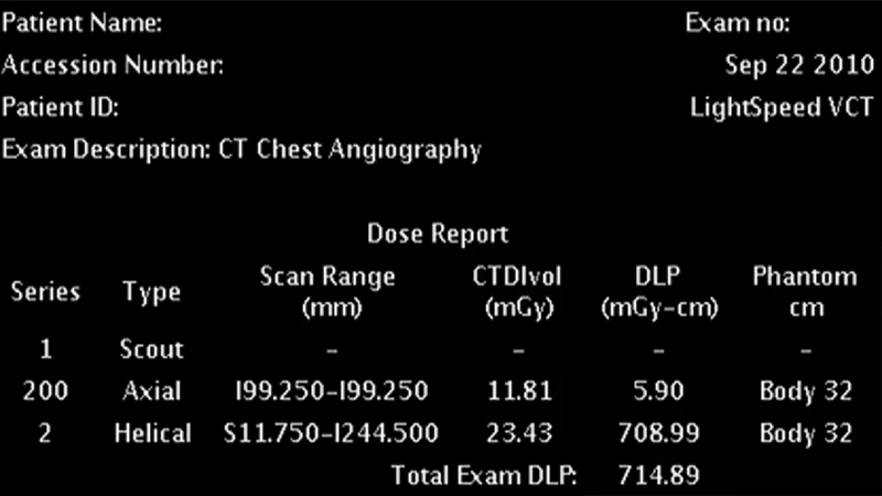
Every modern CT scanner automatically generates a radiation dose report that is stored in the PACS along with the images. This report contains important metrics that technologists and physicists use to monitor and optimize radiation output.
- CTDIvol (CT Dose Index volume): Measured in mGy, this represents the average absorbed dose within the scanned volume. It is a standardized measure of the scanner's radiation output for a specific protocol.
- DLP (Dose Length Product): Measured in mGy-cm, this is the CTDIvol multiplied by the scan length in centimeters. It represents the total radiation energy directed at the patient for the entire exam.
The effective dose (in mSv) is not directly measured but is estimated from the DLP using conversion factors.
Conclusion: The Right Unit for the Right Question
Understanding the different units of radiation measurement helps clarify the conversation around dose. **Gray** measures the raw energy absorbed by a tissue. The **Sievert**, used for both **Equivalent** and **Effective Dose**, is a much better tool for estimating the overall biological risk. By carefully tracking these metrics, imaging departments can ensure they are adhering to the ALARA principle, providing the highest quality of care while minimizing risk to the patient.
Comments