
Common Imaging Artifacts (CT & MRI) and How to Troubleshoot Them
In a perfect world, every medical image would be a flawless representation of the patient's anatomy. In reality, a variety of factors can degrade image quality, creating "artifacts"—features seen in an image that are not present in the actual object. For a technologist, recognizing, understanding, and preventing artifacts is a critical skill for producing diagnostic-quality studies.
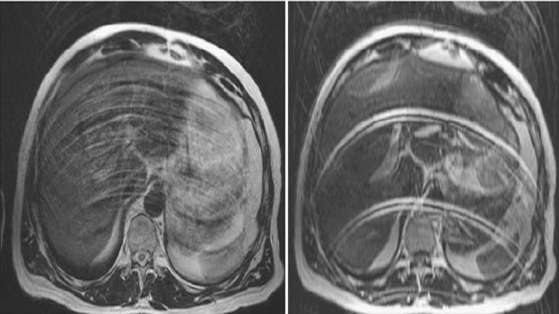
1. Motion Artifact
This is the most common artifact across all imaging modalities. It occurs when the patient moves during the scan acquisition, resulting in blurring, ghosting, or streaking across the image.
- Appearance: Blurry or smeared anatomy, ghost-like repetitions of structures.
- Cause: Voluntary (e.g., patient moving a limb) or involuntary (e.g., breathing, heartbeat, peristalsis) patient movement.
- Solution:
- Clear Communication: Carefully explain the importance of holding still to the patient. Use clear breath-hold instructions.
- Immobilization: Use straps, pads, and cushions to help the patient remain comfortable and still.
- Technique: Use faster imaging sequences when possible. For MRI, techniques like respiratory gating can synchronize the scan with the patient's breathing.
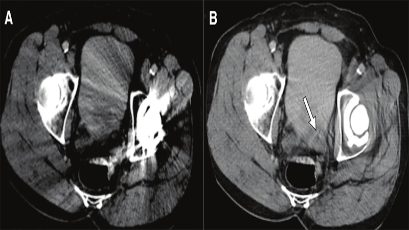
2. Metal Artifact
Metal objects on or inside the patient can severely distort the image, particularly in CT and MRI.
- Appearance (CT): Severe streaking or "starburst" patterns originating from the metal object.
- Appearance (MRI): Large, dark signal voids with bright, distorted edges. The distortion is often much larger than the object itself.
- Cause: High-density metal (like dental fillings, surgical clips, or joint replacements) completely absorbs X-rays in CT. In MRI, ferromagnetic metals distort the local magnetic field.
- Solution:
- Screening: The best solution is prevention. Thoroughly screen patients for removable metal (jewelry, piercings, clothing).
- Positioning: If possible, position the patient so the metal is outside the primary area of interest.
- Software: Modern scanners have dedicated Metal Artifact Reduction Software (MARS) algorithms that can help correct the image during reconstruction.
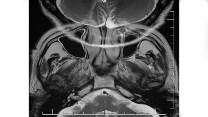
3. Aliasing or "Wrap-Around" Artifact (MRI)
This is a specific MRI artifact that occurs when the Field of View (FOV) is set too small for the body part being scanned. Anatomy that is outside the FOV gets "wrapped around" and appears on the opposite side of the image.
- Appearance: A portion of the anatomy (e.g., the nose in a brain scan, or an arm in an abdomen scan) appears superimposed on the other side of the image.
- Cause: The scanner misinterprets the spatial location of signals coming from outside the prescribed FOV.
- Solution:
- Increase FOV: The simplest fix is to ensure the Field of View is large enough to encompass the entire anatomy being imaged.
- Oversampling/No Phase Wrap: Most modern scanners have built-in software options (often called "oversampling" or "no phase wrap") that can be enabled to prevent this artifact without having to significantly increase the FOV.
Conclusion: The Art of Troubleshooting
Producing high-quality medical images is both a science and an art. While scanners are technologically advanced, they are not fully automatic. A great technologist is a skilled troubleshooter who can anticipate potential problems, recognize artifacts when they occur, and apply the correct techniques to mitigate them. By mastering the control of artifacts, technologists play a direct role in ensuring that radiologists have the clear, accurate images they need to make a confident diagnosis.
Comments