
The Principles of Magnetic Resonance Imaging (MRI)
Magnetic Resonance Imaging (MRI) is a sophisticated medical imaging technique that stands apart from X-ray and CT because it does not use ionizing radiation. Instead, it uses a powerful magnetic field, radio waves, and a computer to create exquisitely detailed images of the body's internal structures. Its greatest strength is its unparalleled ability to visualize soft tissues, making it the gold standard for imaging the brain, spine, joints, and muscles.
The Core Physics: Protons, Pulses, and Signals
The science behind MRI is complex, but it's based on the behavior of hydrogen atoms—which are abundant in the water and fat that make up the human body. Each hydrogen atom's nucleus behaves like a tiny, spinning magnet, referred to as a proton.
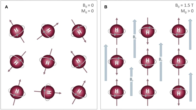
- The Big Magnet (Alignment): Normally, these protons spin in random directions. When a patient is placed inside the strong magnetic field of an MRI scanner (the B0 field), the protons align with that field, much like a crowd of compass needles all pointing north.
- The Radiofrequency (RF) Pulse: Next, the MRI machine sends a radio wave pulse into the patient at a specific frequency. This pulse of energy is absorbed by the protons, knocking them out of alignment.
- The Signal (Relaxation): When the RF pulse is turned off, the protons begin to "relax" and realign with the main magnetic field. As they do, they release the energy they absorbed, emitting a faint radio signal. This signal is detected by a receiver coil in the MRI scanner.
The magic of MRI is that different tissues (like muscle, fat, brain matter, and fluid) relax and release their signals at different speeds. The computer analyzes these timing differences to construct a detailed image.
The Language of MRI: T1 and T2 Weighting
The process of protons relaxing back into alignment occurs in two distinct ways, known as T1 and T2 relaxation. By adjusting the timing of the RF pulses and listening for the signal, technologists can create images that emphasize one type of relaxation over the other. This is called "image weighting."
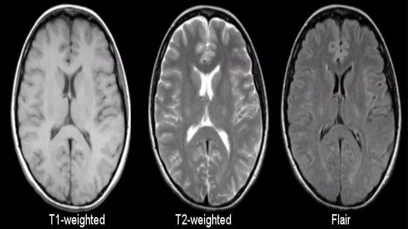
- T1-Weighted Images: In these images, tissues that relax quickly (like fat) appear bright. Tissues that relax slowly (like water and cerebrospinal fluid) appear dark. T1 images are excellent for visualizing normal anatomy and the structure of organs.
- T2-Weighted Images: In these images, tissues that relax slowly (like water and fluid) appear bright. This makes T2 images extremely sensitive for detecting pathology, as many diseases—like tumors, inflammation, and trauma—result in an increase in water content.
Common Applications: Where MRI Excels
Due to its superb soft tissue contrast, MRI is the preferred modality for a wide range of diagnostic tasks:
- Neurology: It is the best tool for examining the brain and spinal cord, detecting tumors, strokes, multiple sclerosis, and herniated discs.
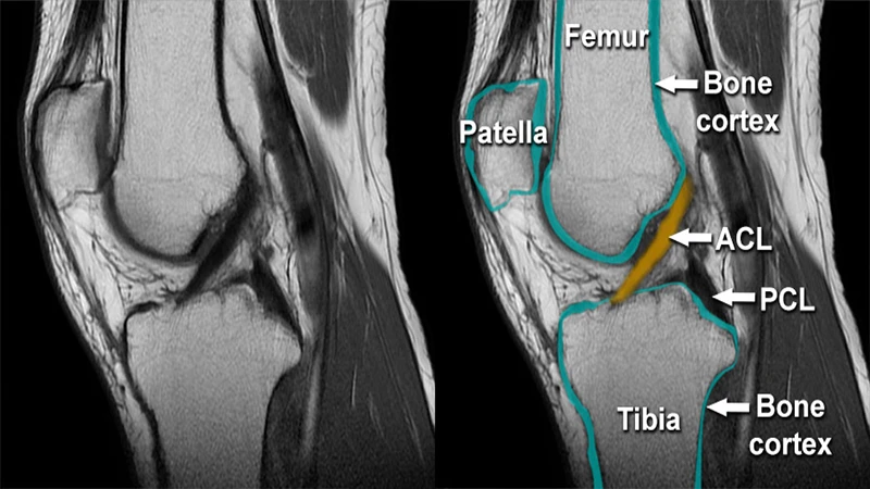
- Musculoskeletal (MSK): MRI is used to diagnose injuries to ligaments, tendons, cartilage, and muscles, such as a torn ACL in the knee or a rotator cuff tear in the shoulder.
- Abdominal & Pelvic Imaging: It provides detailed views of organs like the liver, kidneys, and pancreas, and is frequently used in gynecological and prostate imaging.
Safety is Paramount
The most critical aspect of MRI is safety. The scanner's magnet is incredibly powerful and is **always on**. Before entering the MRI room, all individuals must be thoroughly screened for any metal on or in their body. Ferromagnetic objects can become dangerous projectiles if brought near the magnet.
Key contraindications include pacemakers, certain types of aneurysm clips, and cochlear implants. Patients also experience loud, repetitive knocking sounds during the scan and must remain very still for what can be a lengthy examination (often 30-60 minutes).
Conclusion: Unmatched Detail Without Radiation
Magnetic Resonance Imaging offers a window into the body's soft tissues that no other imaging modality can match. By cleverly manipulating the magnetic properties of atoms already within us, it generates images of stunning detail and diagnostic power. While it is more time-consuming and expensive than CT, its lack of ionizing radiation and superior soft tissue contrast make it an irreplaceable tool in modern medicine.
Comments