X-ray vs. CT Scan: What's the Difference?
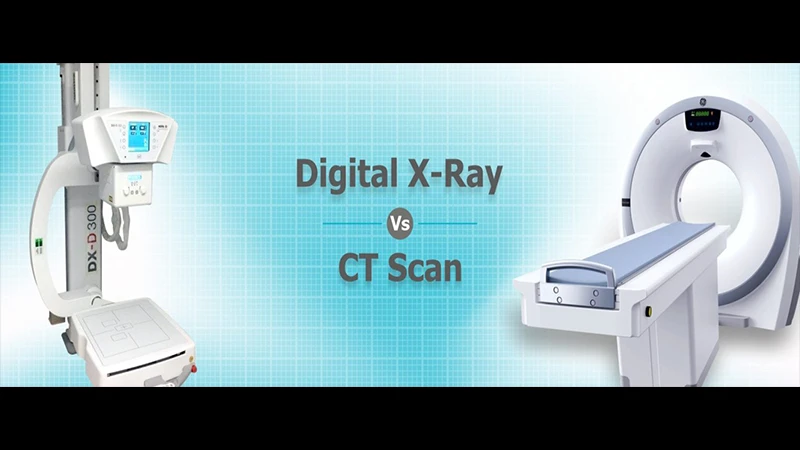
Both X-rays and CT (Computed Tomography) scans are common medical imaging tests that use X-ray radiation to see inside the body. While they are based on the same fundamental technology, they produce very different types of images and are used for different reasons. Think of a standard X-ray as a simple photograph and a CT scan as a highly detailed, 3D model.
The Technology: A Single Picture vs. a 360-Degree View
The biggest difference is how the images are created.
- X-ray: A standard X-ray machine sends a single, stationary beam of radiation through the body to a detector on the other side. This creates one flat, 2D image. It is like casting a shadow on a wall.
- CT Scan: A CT scanner is a much more complex, doughnut-shaped machine. An X-ray source and detector spin rapidly around the patient, taking hundreds of X-ray "slices" from every angle. A powerful computer then reconstructs these slices into detailed, cross-sectional 2D images and can even assemble them into a 3D model.

The Images: A Flat Shadow vs. Detailed Slices
The difference in technology leads to a huge difference in the final images.
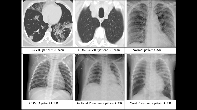
- X-ray Image: An X-ray shows a 2D view where all the internal structures are superimposed on top of each other. For example, on a chest X-ray, the ribs, lungs, heart, and spine are all layered together. This can make it difficult to see small details behind other organs.
- CT Image: A CT scan eliminates this overlap. It provides a clear, cross-sectional view of one "slice" of the body at a time, as if you were looking at a single slice of a loaf of bread. This allows doctors to see the precise size, shape, and location of organs and abnormalities with incredible clarity.

When Are They Used?
The choice between an X-ray and a CT scan depends on what your doctor needs to see.
- X-ray is best for:
- Diagnosing simple bone fractures.
- Screening for lung diseases like pneumonia or a collapsed lung.
- Detecting swallowed foreign objects.
- Screening mammography.
- CT is best for:
- Diagnosing complex fractures or subtle bone injuries.
- Evaluating internal injuries to organs after trauma.
- Detecting and staging cancer, and monitoring its response to treatment.
- Diagnosing blood clots, aneurysms, and other vascular problems (with contrast).
- Investigating the cause of severe, unexplained pain.
Radiation Dose and Time
Because a CT scan takes many more pictures than a standard X-ray, it involves a higher dose of radiation. A chest X-ray has a very low dose, while a chest CT has a dose that is significantly higher, but still considered safe when medically necessary. An X-ray is also much faster, taking only a few seconds, while a CT scan takes a few minutes.
Conclusion: The Right Tool for the Question
An X-ray is a fast, low-dose, and inexpensive tool that is perfect for many common diagnostic questions, especially involving bones and the chest. A CT scan is a more powerful, detailed, and higher-radiation exam that is used for more complex problems, emergency situations, and when a clear, 3D understanding of the anatomy is required. Your doctor will always choose the simplest test that can safely and effectively answer the specific clinical question at hand.


Comments