
Mammography: The Gold Standard in Breast Imaging
Mammography is a specialized medical imaging technique that uses a low-dose X-ray system to see inside the breasts. For decades, it has been the most effective primary tool for the early detection of breast cancer, capable of identifying abnormalities long before they can be felt. Mammography plays a crucial role in both screening asymptomatic women and diagnosing those with symptoms.
How a Mammogram Works: Low Dose, High Detail
A mammography machine is essentially a refined X-ray system designed specifically for breast tissue. It uses a lower dose of radiation than a standard X-ray to produce highly detailed images. The key to a successful mammogram is the ability to differentiate between various types of soft tissue, such as fat, glandular tissue, and potential abnormalities like calcifications or masses.
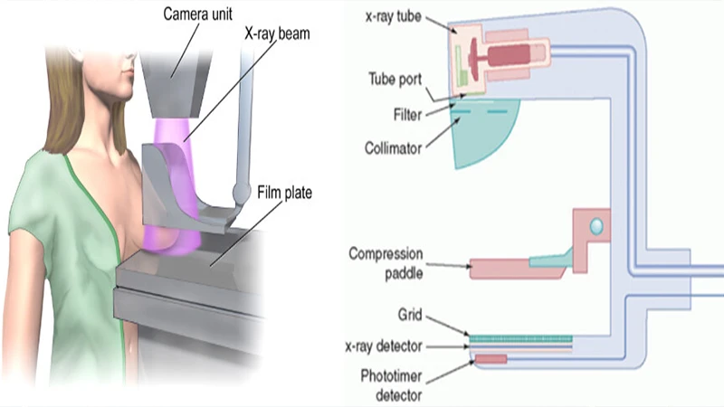
The Critical Role of Compression
One of the most important aspects of mammography is breast compression. A clear plastic paddle is used to gently but firmly flatten the breast tissue. While it can be uncomfortable, compression is essential for several reasons:
- Spreads out tissue: It separates overlapping structures, making it easier to detect small abnormalities that might otherwise be hidden.
- Reduces radiation dose: A thinner breast requires less radiation to produce a high-quality image.
- Prevents motion blur: It holds the breast still, resulting in a sharper image.
Standard screening mammograms typically involve two views of each breast: one from top to bottom (craniocaudal, or CC view) and one from an angle (mediolateral oblique, or MLO view).
Screening vs. Diagnostic Mammography
It's important to distinguish between the two main types of mammograms:
- Screening Mammogram: This is a routine exam for women with no signs or symptoms of breast disease. The goal is to detect cancer at its earliest, most treatable stage.
- Diagnostic Mammogram: This is performed when a woman has a symptom (like a lump) or if a potential abnormality is found on a screening mammogram. It involves additional, specialized views to get a more detailed look at the area of concern.
The Next Generation: 3D Mammography (Tomosynthesis)
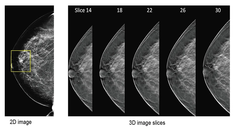
A major advancement in breast imaging is Digital Breast Tomosynthesis, often called 3D mammography. During a tomosynthesis exam, the X-ray tube moves in an arc over the breast, taking multiple images from different angles. A computer then reconstructs these images into a series of thin, one-millimeter slices.
This technique allows the radiologist to examine the breast tissue one layer at a time, similar to flipping through the pages of a book. It helps reduce the problem of overlapping tissue and has been shown to increase the cancer detection rate while reducing the number of "false alarms" or patient callbacks.
Conclusion: A Lifesaving Tool
Mammography remains the cornerstone of breast cancer screening. Through the use of specialized low-dose X-rays and techniques like compression and tomosynthesis, it provides a detailed view of breast tissue that is critical for early detection. Regular screening mammography is one of the most powerful tools available in the fight against breast cancer, significantly improving outcomes and saving lives.
Comments