
An Introduction to Computed Tomography (CT): Seeing the Body in Slices
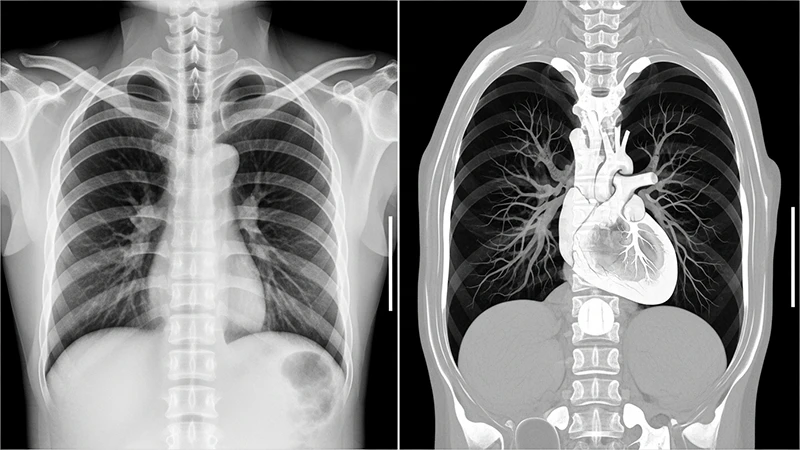
While a conventional X-ray provides a flat, two-dimensional view of the body, Computed Tomography (CT) represents a revolutionary leap forward. By combining X-ray technology with powerful computers, CT scanners produce detailed, cross-sectional images—or "slices"—of the body, allowing us to see inside with unprecedented clarity. It eliminates the overlapping of structures seen in standard radiography, providing a much clearer view of organs, bones, and soft tissues.
The Core Principle: How CT Works
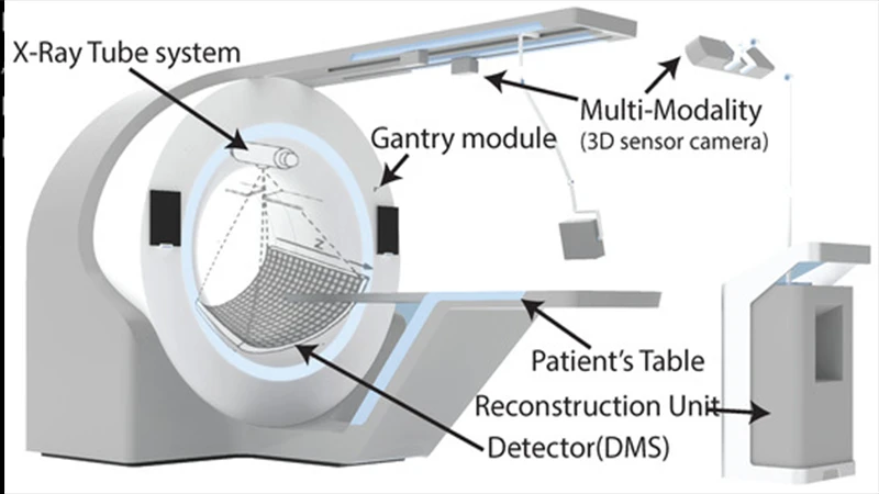
The term "tomography" originates from the Greek words "tomos" (slice) and "graphein" (to write). At its heart, a CT scanner creates a 2D image of a single slice from a 3D object—the patient. It does this using a sophisticated rotating apparatus called a gantry.
Inside the gantry, an X-ray tube and an opposing arc of detectors spin rapidly around the patient. As the gantry rotates, the X-ray tube emits a thin, fan-shaped beam that passes through the body. The detectors on the other side measure the amount of radiation that makes it through from thousands of different angles. This raw data, known as projections, is sent to a computer for processing.
The Language of CT: Hounsfield Units (HU)
The computer's primary task is to perform a complex process called image reconstruction. It uses sophisticated algorithms to translate the thousands of projection measurements into a grid of pixels, where each pixel is assigned a specific shade of gray. This is not a random assignment; each shade corresponds to a calculated tissue density.
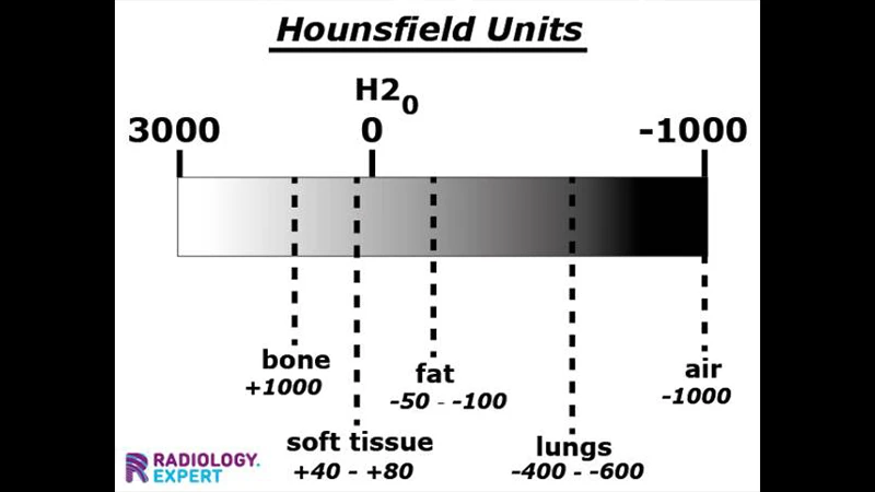
This density is measured on a standardized scale known as **Hounsfield Units (HU)**, named after the engineer Sir Godfrey Hounsfield, one of the principal inventors of the CT scanner. The scale is calibrated based on the density of distilled water.
- Water is defined as 0 HU.
- Tissues denser than water, like bone, absorb more X-rays and have positive HU values.
- Tissues less dense than water, like fat and air, absorb fewer X-rays and have negative HU values.
This numerical precision is what makes CT so powerful for diagnosis. Radiologists can measure the HU of a suspicious finding to help determine if it's a simple cyst (fluid-filled, ~0 HU) or a solid tumor.
Common Applications: When is a CT Scan Used?
CT's speed and detail make it an indispensable tool in many areas of medicine:
- Trauma & Emergency: In emergency rooms, CT is the modality of choice for quickly identifying internal bleeding, organ injury, and complex bone fractures after an accident.
- Oncology (Cancer): CT scans are used to detect tumors, determine their size and location (staging), and monitor how they respond to treatment like chemotherapy.
- Vascular Disease: By injecting an iodine-based contrast agent into the bloodstream (a procedure known as CT Angiography), doctors can visualize blood vessels to diagnose conditions like aneurysms and blockages.
- Neurology: For acute neurological symptoms, CT is often the first test used to diagnose a stroke or bleed in the brain due to its speed.
The Patient Experience
For the patient, a CT scan is a relatively straightforward procedure. You will be asked to lie on a motorized table that gently moves you into and out of the doughnut-shaped gantry. The machine may make whirring and clicking sounds as it operates. To ensure clear images, you may be asked to hold your breath for short periods. Sometimes, an IV contrast agent is administered to make certain tissues and blood vessels stand out more clearly, which may cause a temporary warm sensation throughout the body.
Conclusion: A Powerful Diagnostic Tool
Computed Tomography has fundamentally changed medicine by providing a fast, non-invasive way to look inside the human body. Its ability to produce detailed, cross-sectional images makes it a cornerstone of diagnosis for a vast range of conditions. While CT does involve ionizing radiation, technologists and radiologists are guided by the **ALARA (As Low As Reasonably Achievable)** principle, always using the lowest dose necessary to obtain high-quality diagnostic images. This ensures that the significant benefit of a medically necessary CT scan far outweighs the risk.
Comments